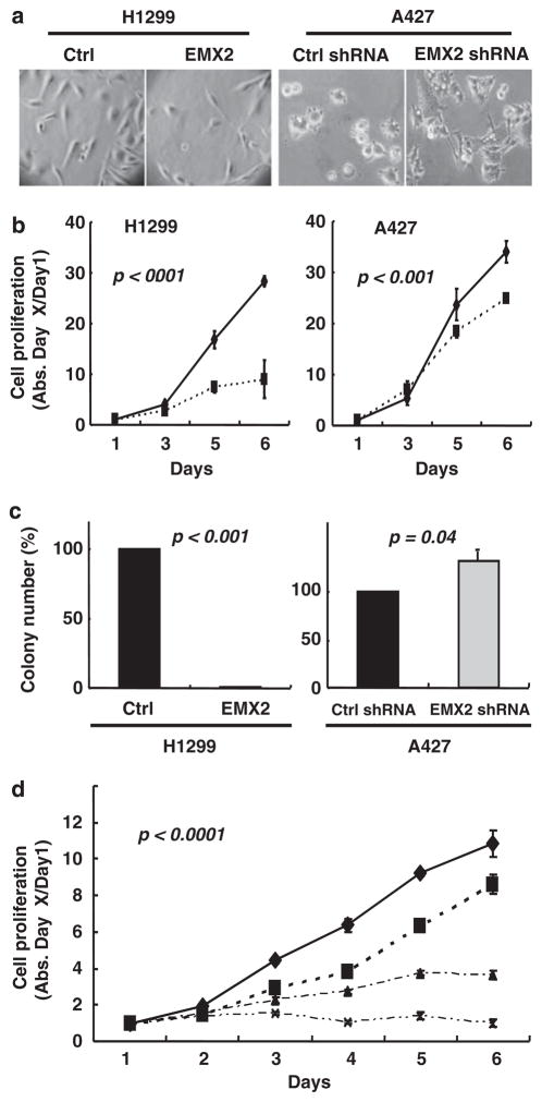
Figure 2.
EMX2 suppressed lung cancer cell proliferation and sensitized lung cancer cells to cisplatin. H1299 cells were transfected with pcDNA 3.1/EMX2 mammalian expression vector (subcloned from pCMV6-XL5/EMX2 vector (Origene, Rockville, MD, USA)). A427 cells were transfected with EMX2 shRNAs (5′-TCAAGCCATTTACCAGGCTTCGGAGGAAG-3′ and 5′-CGG TGGAGAATCGCCACCAAGCAGGCGAG-3′) and non-silencing shRNA (all in pRFP-C-RS vector, Origene). Transfection was done using Lipofectamine2000 (Invitrogen). Transfected cells were re-plated from six-well plates to 10 cm dishes for selection with G418 (500 μg/ml; Invitrogen). Stable transfectants were maintained in regular medium with G418 (300 μg/ml) before analyses. (a) Morphology under light microscope (×40). (b) MTS assay of H1299 cells stably transfected with EMX2 (solid diamonds) and empty pCDNA3.1 vector control (solid squares); and A427 cells stably transfected with EMX2 shRNA (solid squares) and non-silencing shRNA control (solid diamonds). Controls were set as 100%. Proliferation assay was performed by plating the stably transfected cells in 96-well plates at a density of 500–1000 cells/well in 100 μl of G418 culture medium. Medium was changed every day. Cell viability was evaluated in triplicate by CellTiter 96 AQueous (Promega, Madison, WI, USA). (c) Colony formation assay. In all, 500 individual stably transfected cells were seeded in 10 cm dishes and cultured for 10 days. Colonies were then fixed by 10% formalin, stained with 0.5% crystal violet and counted. (d) Synergistic effect between EMX2 and cisplatin in H1299. Diamonds, squares, triangles and crosses are treatments of control vector alone, control vector+cisplatin (0.3 ng/ml), EMX2 cDNA alone and EMX2 cDNA+cisplatin (0.3 ng/ml), respectively. All results are means±s.d. (error bars). Differences between groups were compared with a two-sided Student’s t-test. A P-value of ≤0.05 was considered to be significant.

