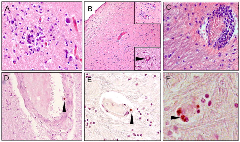
Figure 2.

Neuropathological alterations in hypothalamic sections from the brains of HIVE+ patients. A) microglial nodule at the grey/white matter junction; B) low magnification of the periventricular region of the 3rd ventricle. The top right corner inset shows inflammatory cell infiltration surrounding small vessels. The lower right corner inset shows a multinucleated giant cell (arrowhead); C) perivascular cuffing by infiltrating macrophages and lymphocytes with expansion of the perivascular space in the white matter; D) p24 immunoreactive macrophage in the vascular space of a small artery of the blood brain barrier (arrowhead); E) p24 immunoreactive macrophage adjacent to a small vessel (arrowhead); F) higher magnification of 2-3 p24 immunoreactive cells (arrowhead) associated with a disrupted microvessel. Magnification for A, C, D, E is 40×, B is 10× (insets are 40×), F is 100×.
