
FIG. 11.
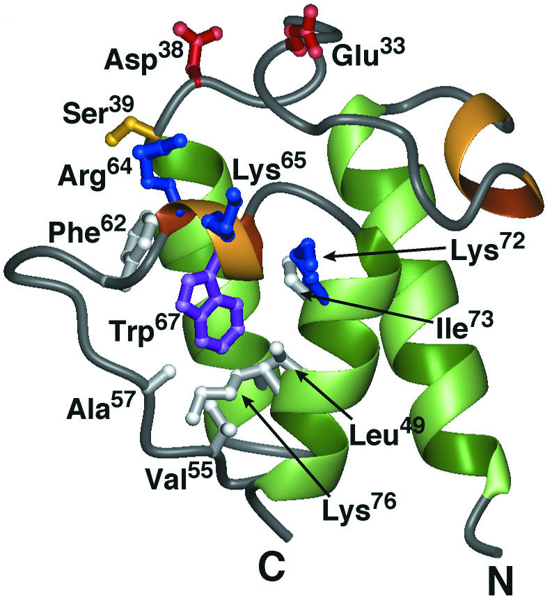
Ribbon diagram of the minimized average structure of apo-Dcp (PDP entry 1HQB). Residues shown in white bury the Trp67 side chain (purple) in the hydrophobic core. Other key residues include the conserved Glu33 and Asp38(red) and Ser39 (yellow), as well as a cluster of basic residues (blue) proximal to the phosphopantetheine attachment site (Arg64, Lys65, and Lys72). Reprinted from reference 485 with permission of the publisher.