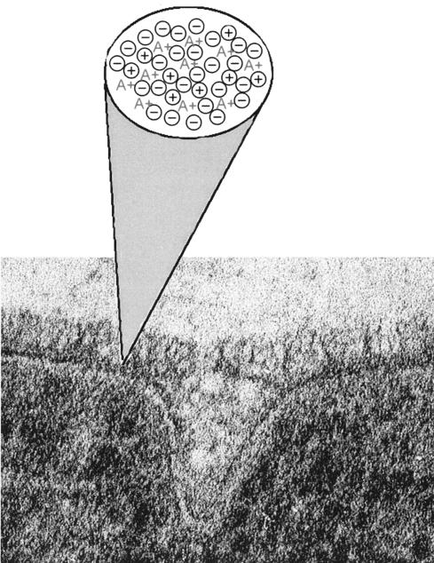
FIG. 20.
Continuum of ionic charge. A high-magnification, freeze-substituted image of the septal region of an exponentially growing B. subtilis 168 cell is shown. The tripartite structure of the wall shows the fibrous nature of the outer layer. The electron photomicrograph is reprinted from reference 189 with permission. (A+) represents the d-alanyl esters of TAs, ⊕ represents mobile cations and other fixed cationic functions on peptidoglycan, and ⊖ represents the phosphodiester anionic linkages of TAs and anionic groups of peptidoglycan.

