
Figure 1. Near-infrared spectroscopy (NIRS) monitoring of tumor blood oxygenation in response to CA4P.
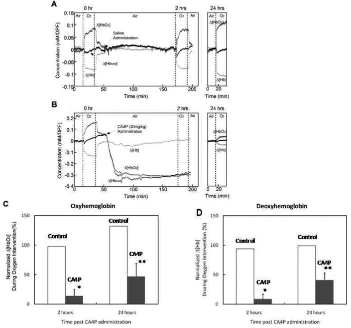
A) For a control tumor, real-time NIRS revealed stable baseline and significant increase in oxyhemoglobin (Δ[HbO2]) observed immediately after switching from air to oxygen breathing (p < 0.0001). Mirrored by significantly decreased deoxyhemoglobin (Δ[Hb]; p < 0.0001). Saline injection caused no marked change and reproducible oxygen response was observed 2 h and 24 h later. B) Following CA4P (30 mg/kg) administration, both Δ[HbO2] and Δ[Hbtotal] dropped rapidly with continuing decline for about 25 min, which remained at the lowest level 2 h post CA4P (p < 0.001). At this time, oxygen breathing no longer produced a significant increase in Δ[HbO2]. However, 24 h later, a small, but significant increase in Δ[HbO2] was evident (p < 0.005). ANOVA Fisher's PLSD was used for testing statistic significance. C) Normalized oxyhemoglobin response to oxygen breathing at 2 h and 24 h was compared in the CA4P treated tumors (n = 3) and saline controls (n = 2). The amplitude change in Δ[HbO2] was normalized to the baseline oxygen response, showing only 14 ± 11 (sd)% increase in Δ[HbO2] at 2 h, but recovery to 47 ± 22 (sd)% of the baseline level 24 h later in CA4P treated tumors. D) Similarly, the normalized mean Δ[Hb] was plotted for CA4P or saline treated groups of tumors. * p < 0.05 to pretreatment, ** p < 0.05 to 2 h; paired Student's t-test.