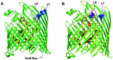
FIG. 6.
X-ray crystallographic structures of the ferric citrate receptor, FecA, of E. coli. (A) Side view of the unliganded FecA. The “plug” domain inside the β-barrel is shown in orange. At its N-terminal end, the short sequence comprising the Ton box is shown in light blue. Loops 7 and 8 are shown in deep blue and mauve, respectively. (B) Liganded FecA. On binding of the ferric citrate (with two large blue balls near the top indicating the two iron atoms, and citrate molecules in stick diagrams), large displacements are seen at the N-terminal end of the plug domain, where residues 80 through 95 (including the Ton box of residues 80 to 84) become disordered and invisible. Loops 7 and 8 also undergo large conformational changes, with the loss of part of the helical structure in loop 7. The diagrams are based on PDB files 1KMO and 1KMP.

