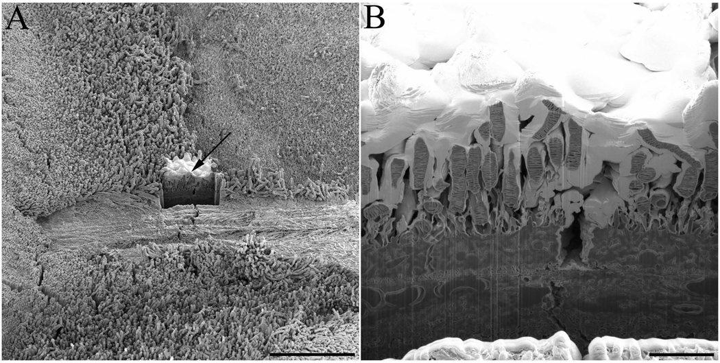
Fig. 2. Experimental set-up for photoreceptor observation with FIB-SEM technology.
(A) Once an area of interest in critical point dried sample of wt mouse retina was selected it was protected by application of a thin layer of platinum inside the FIB-SEM (indicated by arrow), a trench was created by application of an ion beam cleaning cross section. (B) A close-up view of the created trench reveals interior elements of rod photoreceptors that can be imaged by the electron beam. Scale bars in panels A and B are 50 and 5 µm, respectively.

