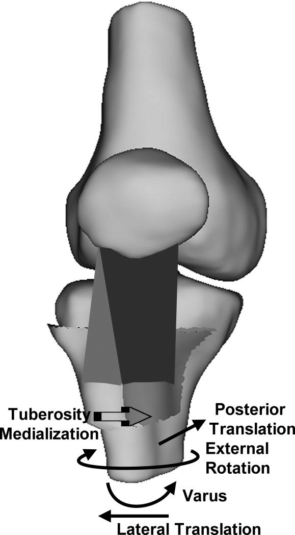
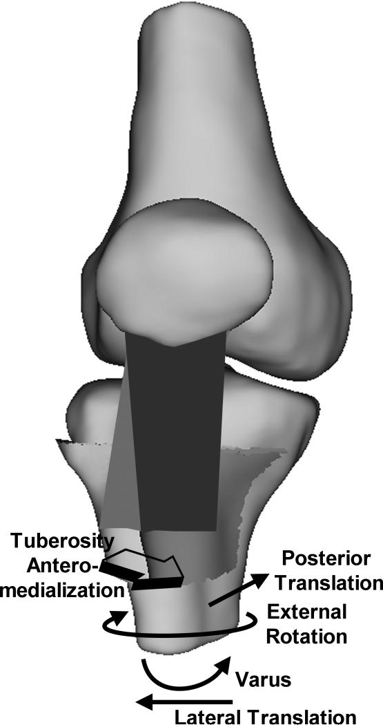
Fig. 6.
Schematic diagram illustrating the directions of the changes in the translations and rotations of the tibia with respect to the femur following medialization (A) and anteromedialization (B) of the tibial tuberosity. The trends were similar for both types of realignment, although the posterior translation tended to be larger for anteromedialization than medialization. The tuberosity is shown in the pre-operative position on the tibia with an elevated TT-TG distance, with the direction of tuberosity realignment indicated.


