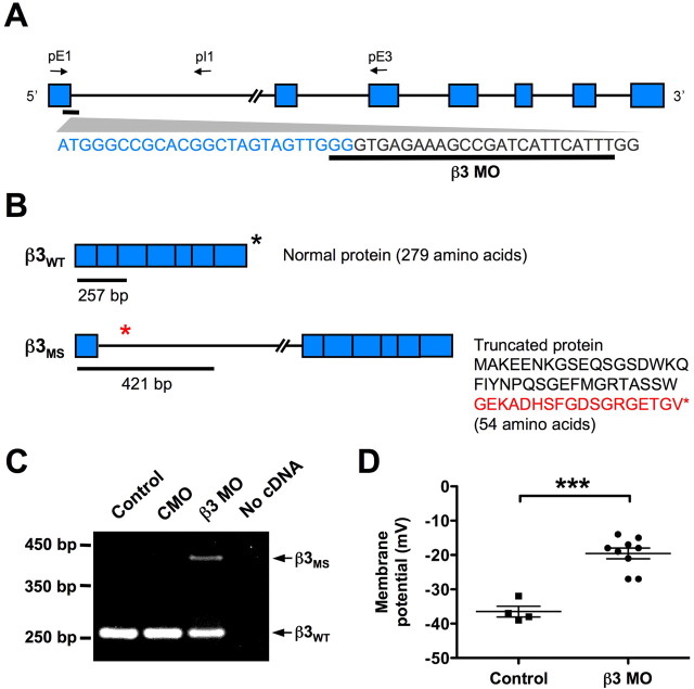
Figure 3.
A splice-blocking morpholino (β3 MO) disrupts normal splicing of the Na+, K+-ATPase β3 subunit in X. tropicalis and alters the membrane potential of embryonic spinal neurons. A, Schematic of β3 unspliced RNA and the β3 MO target sequence. The blue boxes indicate exons, connecting lines represent introns, and arrows show approximate locations of primers used for RT-PCR. B, Predicted splicing of β3 mRNA in wild-type embryos and embryos after β3 MO treatment. Primers shown in A amplify a 257 bp fragment in wild-type β3 mRNA (β3WT) (top) and a 421 bp fragment in misspliced β3 mRNA (β3MS) (bottom). β3WT generates a 279 aa protein, whereas conceptual translation of the predicted β3MS produces a 54 aa protein. The last 18 aa of this truncated protein (red text) are translated from the first intron. The asterisks represent the stop codon location in each transcript. C, RT-PCR of stage 15/16 β3 MO-treated embryos using primers shown in A yields predicted spliced and misspliced products schematized in B. Control uninjected and CMO-treated embryos do not produce the β3MS transcript. D, Intracellular recordings of stage 21/22 ventral spinal neurons from embryos injected with β3 MO in one blastomere at the two-cell stage. Neurons on the β3 MO-treated side display significantly more depolarized membrane potentials than neurons on the control side (***p < 0.0001). Each point represents a recording from a single neuron. The horizontal lines represent the mean ± SEM.

