
Figure 3. Feedforward GABAA-mediated inhibition activated via TA pathway stimulation spatially restricts evoked EPSPs to the distal dendrites of the CA1 pyramidal neurons.
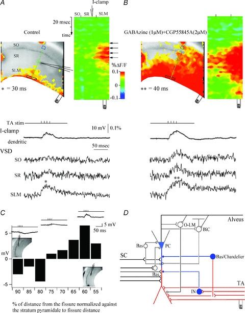
A, control. A snapshot of activation at 30 ms of the VSD responses of evoked EPSPs (denoted by asterisk) to stimulation in stratum lacunosum moleculare (left) and the activation profile (right) generated from the raster line scan along the path of interest (green line). The location of the patch recording electrode is denoted by the electrode graphic. Top trace (I-clamp, current clamp recording), whole-cell recording trace from the apical dendrite of a CA1 pyramidal cell in stratum radiatum. VSD SO, SR, and SLM are the local VSD signals quantified from regions of interest in stratum oriens (blue box), stratum radiatum (green box), and stratum lacunosum moleculare (black box). Note that the TA-evoked EPSP is spatially restricted to the extreme distal dendrites of CA1 pyramidal neurons. B, effects of the GABAA and GABAB antagonists gabazine (1 μm) and CGP 55845A (2 μm). Left, snapshot at 40 ms. Note that GABAergic inhibition blockade results in loss of spatial segregation of the TA EPSPs in stratum lacunosum moleculare and significant propagation of TA EPSPs to stratum radiatum and stratum oriens. C, plot of dendritic whole-cell recording responses along the dendrites of the CA1 pyramidal neuron, as a function of the normalized distance of the patch electrode from the hippocampal fissure to stratum pyramidale. Insets show the relative current-clamp dendritic specimen records and locations of the patch electrode. Note that TA-evoked IPSPs are prevalent in recordings close to the cell somata, whereas EPSPs are recorded from more distal dendritic sites. D, CA1 schematic showing the response to TA stimulation. Red represents excitation and blue represents inhibition. SC, Schaffer collateral; TA, temporoammonic pathway; PC, pyramidal cell; O-LM, oriens–lacunosum moleculare interneuron; BiC, bistratified cell; Bas, basket cell; Chandelier, chandelier cell; IN, interneuron. From Ang et al. (2005), with permission from the Society for Neuroscience.