
Figure 1. Location and electrophysiology of mouse Im neurons.
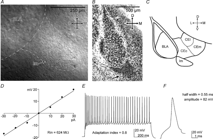
A–C, the Im nucleus can be identified as a cluster of densely packed cells, located ventro-medially to the BLA. A, identification of Im nucleus on mouse coronal slice with IR-DIC microscopy (10×/0.3 NA water-immersion objective). B, anti-NeuN staining revealing the Im. C, scheme of the amygdala depicting the position and the size of the Im at −0.9 bregma level. D–F, passive and active electrophysiological responses of a representative Im cell. D, the I–V plot used to measure the cell Rin. E, sustained firing pattern in response to a depolarising current stimulus (150 pA, 1 s). F, action potential evoked by a short depolarising current pulse (100 pA, 3 ms). Abbreviations: D, dorsal; L, lateral; M, medial; V, ventral; BLA, basolateral complex; CEc, central nucleus, capsular; CEl, central nucleus, lateral; CEm, central nucleus, medial; Im, main intercalated nucleus; Rin, input resistance.