
Figure 3. Hyperpolarising effect of dopamine on the Im cells.
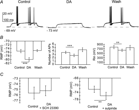
A, response of Im cell to superimposed depolarising current pulses (60 pA, 200 ms) during control, dopamine bath application (DA, 30 μm) and washout. B, average (±s.e.m.) effect of dopamine on the resting membrane potential (RMP, n = 20), number of action potentials (APs; n = 20) and input resistance (Rin; n = 8). Comparison between control and DA bath application (***P < 0.001; **P < 0.01). C, average (±s.e.m.) effect of dopamine on the resting membrane potential (RMP) in the presence of the D1R antagonist SCH 23390 hydrochloride (5 μm, P > 0.1, n = 4) or the D2R antagonist sulpiride (5 μm, **P < 0. 01, n = 4).