
Figure 7. eEPSCs recorded in the Im – occlusion test.
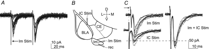
A, voltage clamp recording showing paired pulse facilitation of EPSC evoked by Im stimulation. Individual eIPSCs are shown in grey; the average of 10 traces is shown in black. B, scheme of the amygdala depicting the location of the stimulating and recording electrodes. C, evoked EPSCs recorded in the presence of bicuculline (5 μm), individual eEPSCs are shown in grey; the average of 10 traces is shown in black. Left panel: representative EPSCs evoked by the independent stimulation of Im (peak amplitude = 100.9 pA) and IC (peak amplitude = 54.7 pA). The arrow next to the top traces indicates the time shift applied online to achieve simultaneous stimulation of IC and Im. Right panel: simultaneous activation of Im and IC (peak amplitude = 172.9 pA). In this representative example, the response evoked by the paired stimuli of Im and IC was 111% of the summed individual eEPSCs. Abbreviations: D, dorsal; L, lateral; M, medial; V, ventral; BLA, basolateral complex; CE, central nucleus; IC, intermediate capsula; Im, main intercalated nucleus; Rec., recording electrode, Stim., stimulating electrode.