
Figure 8. AMPA/NMDA receptors mediated eEPSCs recorded in the Im.
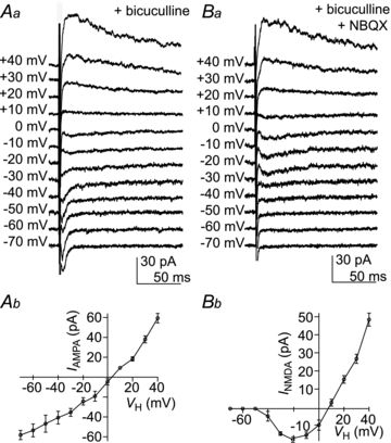
Voltage clamp recording after Im stimulation at VH ranging from −70 mV to + 40 mV. Aa, AMPA receptor-mediated synaptic currents in the presence of bicuculline (30 μm). Ab, plot of the peak amplitude of AMPA receptor-mediated eEPSCs against VH with the standard error indicated. In this representative example, the EPSCs reversal potential was 3.4 mV. Ba, NMDA receptor-mediated eEPSCs in the presence of bicuculline (30 μm) and NBQX (50 μm). Bb, plot of the peak amplitude of NMDA receptor-mediated eEPSCs against VH with the standard error indicated. In this representative example, the eEPSCs reversal potential was 8.1 mV.