
Figure 5. TBOA-induced slow oscillations in mitral cells have altered shape in OCAM knockouts.
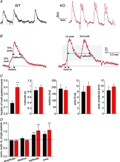
A, application of the glutamate uptake blocker TBOA evoked ongoing slow oscillations of mitral cells. These slow oscillations originate in the glomerular layer because mitral cells with severed apical dendrites show no oscillations (not shown; Schoppa & Westbrook, 2001). Note the presence of frequent multi-peaked oscillations in the knockout. B, we used the following parameters to analyse the shape of individual events: peak amplitude, area, rise time and half-width. For multi-peaked events, a second peak was considered present if the minimum between peaks occurred in the grey area (right panel, see Methods). C, bar graphs depicting mean values for slow oscillations in wild-type (n = 13, black) and knockout (n = 14, red) mitral cells. There was a 3-fold increase in multi-peaked oscillations in the knockout. Membrane potentials were −63.9 ± 1.9 mV and −63.0 ± 1.5 mV for wild-type and knockout respectively. 76 ± 9 and 70 ± 8 events per cell were analysed for wild-type and knockout, respectively. D, for each cell, the parameters of single- and multi-peaked oscillations were normalized to the average values for single-peaked oscillations. The area and half-width was larger for multi-peaked events in the knockout as well as for the occasional multi-peaked events in wild-type cells (paired t test).