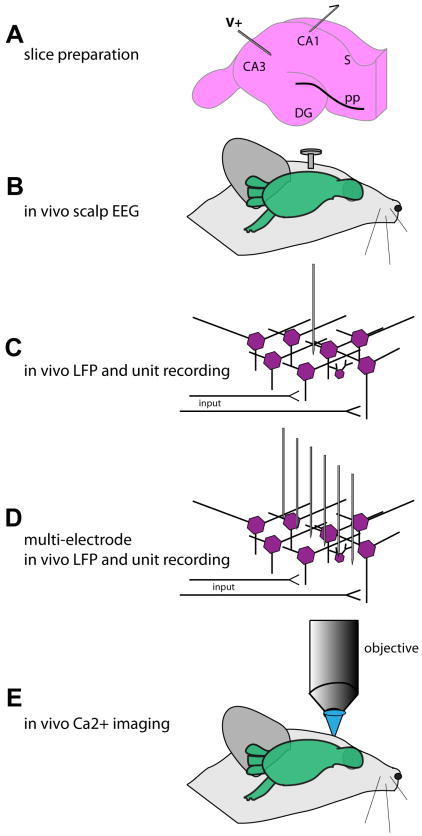
Figure 3. Physiological methods in the study of AD.
A wealth of methods are available in the study of neural circuits in AD models, yet each with its unique strengths and weaknesses. The most common tool in AD model physiology is the hippocampal slice preparation (A) which, while allowing precise recording sites and pharmacological interventions, is in an isolated neural circuit far disconnected from possible behavioral read-outs. Allowing simultaneous physiological and behavioral recordings are the methods depicted in B-E. Whereas in vivo scalp EEG (electroencephalography) presents with relative ease of data collection in comparison to the more precise methods in C, D, and E, it lacks spatial resolution. In contrast, whereas spatially and temporally precise methods shown in C, D, and E allow high levels of resolution, the data collection and analysis can be very time consuming, making the task to interpret large populations of data exceptionally time consuming. LFP = local field potential.

