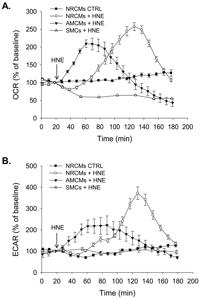
Figure 1. Bioenergetic responses of different cardiovascular cells to electrophile stress.
Extracellular flux analysis of oxygen consumption and glycolysis in neonatal rat cardiomyocytes (NRCMs), adult mouse cardiomyocytes (AMCMs), and rat aortic smooth muscle cells (SMCs). Three baseline measurements of oxygen consumption rate (OCR; panel A) or extracellular acidification rate (ECAR; panel B) were recorded from isolated cardiomyocytes or SMC. Vehicle (DMEM) or HNE was then injected to a final concentration of 20 µM and the measurements then continued for the indicated times. n = 3–5 per group; Repeated measure one-way ANOVA indicated that the response of the groups in panels A and B to HNE differed with respect to time (p<0.05).

