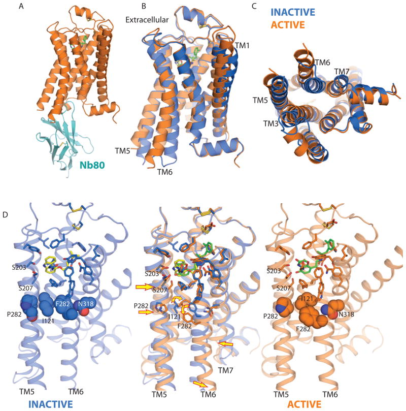
Figure 3.
Active and inactive states of the β2AR. A, β2AR-Nb80 complex. CDR3 of Nb80 projects into the cytoplasmic core of the protein. B, Side view of superimposed active (orange) and inactive (blue) conformations of the β2AR. C. Cytoplasmic view of superimposed active and inactive states. D, Binding pocket of active and inactive states of the β2AR are shown separately (outer panels) and overlapped (center). Small changes in the ligand binding pocket lead to rearrangement of the conserved amino acids (Pro 211, Ile 121, Phe 282, and Asn 318). These changes are indicated by arrows in the center panel. Carazolol is shown with yellow carbons and the agonist BI-167107 is shown with green carbons.

