
Figure 2. Radiation induced NF-κB in colorectal cancer cells.
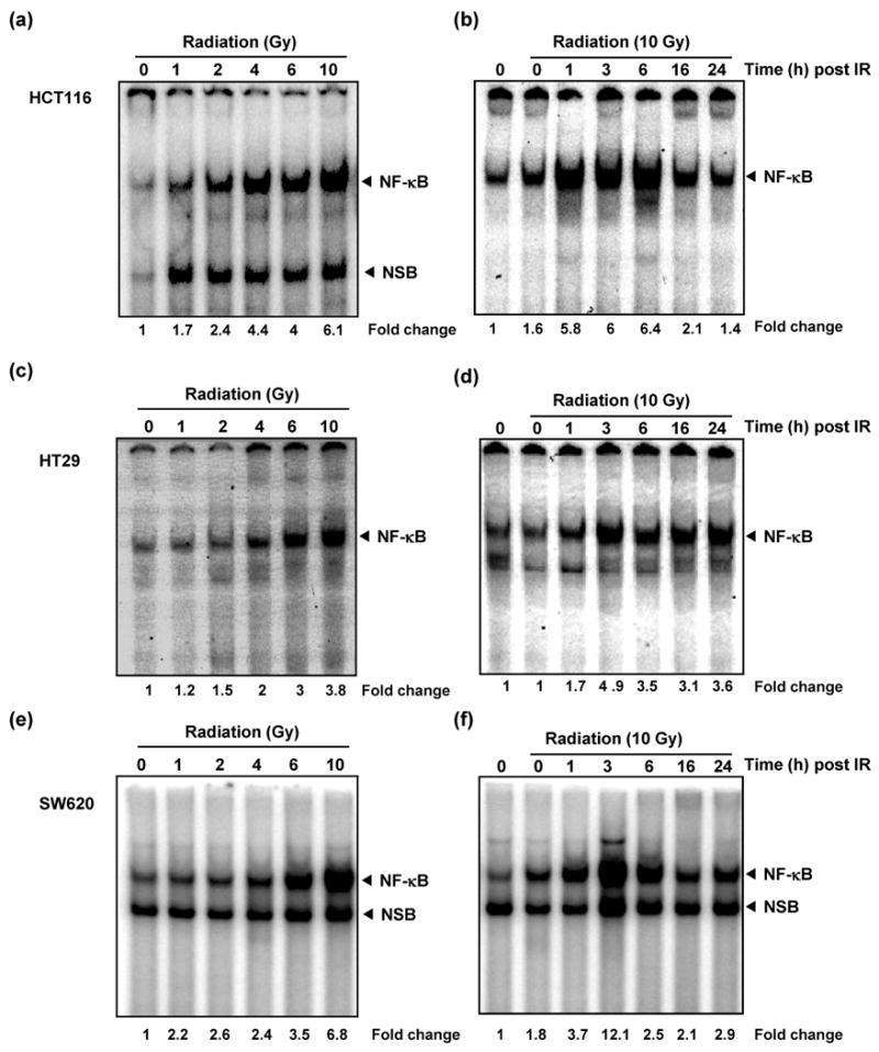
Dose dependent increase in NF-κB activity was observed in HCT116 (a), HT29 (c), and SW620 (e) cells. Colon cancer cells (2 × 10⁵/mL) were exposed to graded doses of radiation and harvested 3 h after irradiation. Nuclear extracts were probed for NF-κB activation by EMSA. The time course of radiation-induced NF-κB activation after 10 Gy irradiation in HCT116 (b), HT29 (d), and SW620 (f). Cells (2 × 10⁵ /mL) were exposed to radiation, harvested post-radiation at indicated time points, and probed for NF-κB. Activation of NF-κB was expressed as fold increase of DNA-bound protein over untreated controls (basal levels). The band labeled NF-κB represents the p65:p50 heterodimer, the predominant functionally active NF-κB dimer whereas the lower band is a non-specific band formed by the inactive NF-κB homodimer (p50:p50)