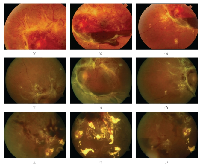
Figure 5.
(a)–(c) Color photographs before intravitreal bevacizumab. Severe proliferative diabetic retinopathy with abundant fibrovascular tissue and subhyaloid hemorrhage. The retina is attached and best-corrected visual acuity (BCVA) is 20/80. (d)–(f) Color photographs 10 days after 2.5 mg of intravitreal bevacizumab demonstrating dense fibrous tissue contraction, and tractional retinal detachment with macular involvement. BCVA is hand motions at 2 meters. (g)–(i) Same eye, eight days after vitrectomy. The retina is reattached and best-corrected visual acuity (BCVA) is 20/120 with silicone oil tamponade.

