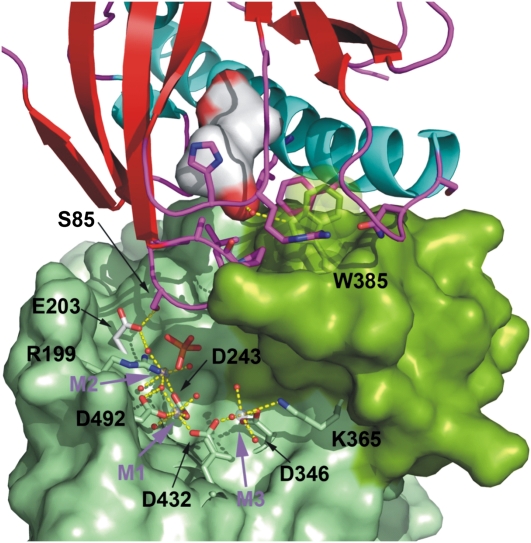
Figure 2.
The PYR1 β3/β4 loop docks at the catalytic site of HAB1. The ABA-bound PYR1 receptor is shown as in Figure 1. The accessible surface of the HAB1 phosphatase is depicted in light green with the flap subdomain containing Trp-385 in dark green. Residues coordinating the three metal ions at the catalytic site were excluded in the calculation of the molecular surface and are depicted as stick models. The water molecules involved in metal coordination are depicted as red spheres. The human PP2Cα structure (not shown), which contains a phosphate ion (shown as stick model) in the active site, was superposed on HAB1 to transfer the position of the phosphate ion into the catalytic site of HAB1.

