Figure 7.
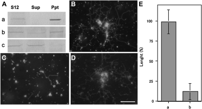
Effects of myosin immunodepletion from the S12 fraction on ER tubule formation. A, Immunodepletion of BY-2 myosin VIII-1 (a) or 175-kD myosin (b) from the S12 fraction by myosin H chain-specific antibodies. The S12 fraction (S12) was mixed with each antibody and then with protein A beads. After centrifugation, the supernatant (Sup) and pellet (Ppt) were recovered and then subjected to immunoblotting with the anti-ATM1 antibody (a) or 175-kD myosin H chain-specific antibodies (b). As a control, the S12 fraction was mixed only with protein A beads and then immunoblotting was carried out using a 175-kD myosin H chain-specific antibody (c). B to D, ER tubule formation in the supernatant shown in Aa, Ab, and Ac, respectively, in the presence of 2 mm ATP, 0.5 mm GTP, and 2.5 μg mL−1 F-actin. Bar = 10 μm. E, Columns a and b show the length of ER tubules formed in the supernatant shown in Aa and Ab, respectively. The length is shown as a relative value (%) to that of the control, and means ± sd from three separate experiments are plotted.

