Figure 1.
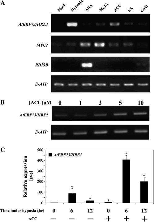
Effects of abiotic stresses and hormones on AtERF73/HRE1 expression. A, RT-PCR analysis of AtERF73/HRE1 expression. The mRNA levels of AtERF73/HRE1, MYC2, and RD29B in roots of 14-d-old seedlings were determined after mock treatment (lane 1) and after treatment with hypoxia (lane 2), 5 μm ABA (lane 3), 50 μm MeJA (lane 4), 5 μm ACC (lane 5), 50 μm SA (lane 6), or cold (4°C; lane 7) for 6 h. β-ATP was used as a control. Experiments were carried out in triplicate with similar results. B, RT-PCR analysis of the effects of ACC. mRNA levels of AtERF73/HRE1 in 14-d-old seedlings were determined after treatment with 0, 1, 3, 5, or 10 μm ACC for 6 h. C, Quantitative RT-PCR analysis of the effects of combined ACC and hypoxic treatment. Levels of AtERF73/HRE1 mRNA in 14-d-old seedlings were determined after treatment with hypoxia for 6 or 12 h in the presence (+) or absence (−) of 5 μm ACC, respectively. The relative amounts of transcripts were calculated and normalized with β-ATP mRNA. Values represent means ± sd from three biologically independent experiments. * P < 0.05 versus the wild type (one-way ANOVA with Dunnett’s t test).

