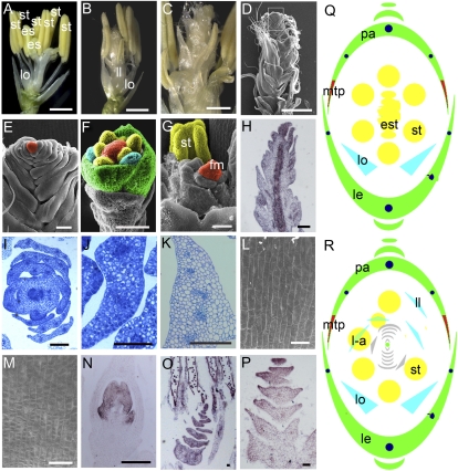
Figure 4.
Flower phenotype of dl-sup6 osmads3-4. A, One dl-sup6 flower showing ectopic stamens in the center. B, One dl-sup6 osmads3-4 flower showing the phenotypes in the second and third whorls similar to osmads3-4. C, Closeup of one dl-sup6 osmads3-4 flower showing mosaic organs and indeterminate organs in the center. D, SEM observation of one dl-sup6 osmads3-4 flower showing the supernumerary whorls of indeterminate undifferentiated organs in the floral center. E, Closeup of D. F, SEM observation of one dl-sup6 osmads3-4 flower at stage Sp6. G, SEM observation of one dl-sup6 osmads3-4 flower after stage Sp7 showing ectopic organs and indeterminate meristem. H, Expression pattern of OSH1 in the dl-sup6 osmads3-4 flower at stage Sp8. I and J, Transverse sections of one dl-sup6 osmads3-4 flower showing the ectopic organs in the flower center. K, Transverse section of one wild-type lodicule. L and M, SEM analysis of epidermal cells of osmads3-4 du-sup6 ectopic organs and wild-type lodicules, respectively. N, Expression of SPW1 in wild-type lodicules and stamens. O, Transcripts of SPW1 detectable in lodicule-like organs of one dl-sup6 osmads3-4 flower center. P, OsMADS15 is expressed in lodicule-like organs in one dl-sup6 osmads3-4 flower center. Q and R, Floral diagrams of dl-sup6 (Q) and osmads3-4 dl-sup6 (R). est, Ectopic stamen; fm, floral meristem; l-a, lodicule-anther mosaic organs; ll, lodicule-like structure; lo, lodicules; mtp, marginal tissue of the palea; st, stamen. Bars = 1 mm in A, B, and D; 500 μm in C; 100 μm in E, H to K, and N to P; 50 μm in F; and 20 μm in L and M.

