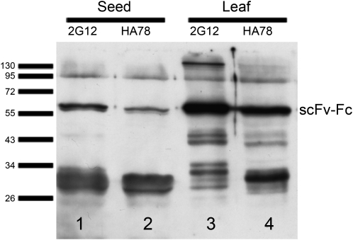
Figure 3.
Immunodetection of 35S-driven scFv-Fcs extracted from seeds and leaves. Nine microliters of crude seed extract and 5 μL of crude leaf extract (corresponding to 90 μg of seeds and 375 μg of leaf material) were separated by SDS-PAGE, blotted on a nitrocellulose membrane, and detected with goat anti-human IgG (H+L) HRP conjugate (Promega; W403B) and a chemiluminescent substrate (SuperSignal West Pico Chemiluminescent Substrate; Pierce). Film exposure for 30 s reveals scFv-Fc bands as well as degradation products. Bars indicate the height of marker bands (in kD).

