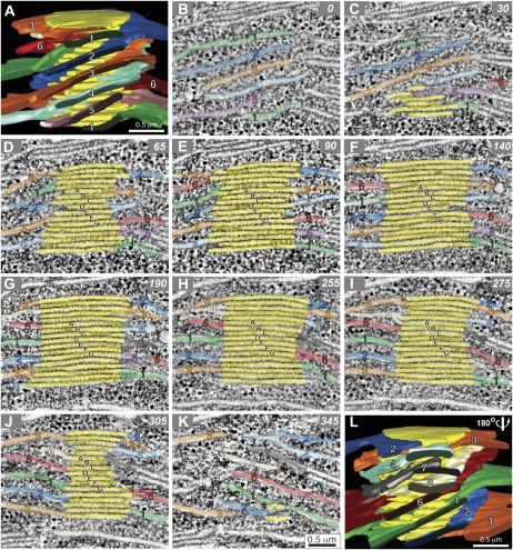
Figure 3.
Serial tomographic slice images through a grana thylakoid stack and reconstructed models of the stack. A is a tomographic model of a grana stack. Grana thylakoids are colored yellow, and interconnecting stoma thylakoids are colored and numbered so their position can be tracked in the serial tomographic slices B to K. B to K show serial 2.2-nm tomographic slice images at specific z intervals through the grana stack.

