Figure 8.
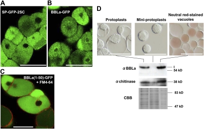
Subcellular localization of BBLa protein in cultured tobacco cells. A, Full-length BBLa protein was fused at its C terminus to GFP and stably expressed in cultured BY-2 cells. Fluorescence was observed with a laser scanning confocal microscope. B, Confocal image of BY-2 cells expressing SP-GFP-2SC, which had been shown to be located in the vacuoles (Mitsuhashi et al., 2000). C, An N-terminal 50-amino acid fragment of BBLa was fused to GFP and stably expressed in the tobacco cells. The transformed tobacco cells were pulse labeled by the fluorescent dye FM4-64 and inspected 10 h later when the dye had been shown to primarily label the vacuolar membrane (Shoji et al., 2009). Bars = 50 μm. D, Immunoblots of subcellular fractions. Vacuoles and cytoplasm-rich miniprotoplasts were prepared from protoplasts of a BBLa-overexpressing tobacco cell line by Percoll gradient centrifugation. Vacuoles were stained with a 10 μg mL−1 neutral red solution for 10 min. Immunoblots probed with antisera against BBL and class I chitinase (vacuolar resident protein) are shown, together with a Coomassie Brilliant Blue (CBB)-stained gel as a loading control. Bars = 100 μm. [See online article for color version of this figure.]

