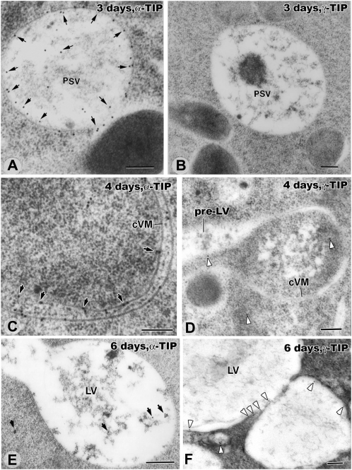
Figure 8.
Immunogold localization of α-TIP and γ-TIP in the tonoplast membranes of inner cortex cells in 3-, 4-, and 6-d seedlings. Note that, unlike in Figures 1, 4, 6, and 7, the samples have not been stained with osmium and that, for this reason, the PSVs are not darkly stained. A and B, PSVs of a 3-d plant whose tonoplast membranes are strongly labeled (arrows) with anti-α-TIP antibodies (A) but not anti-γ-TIP antibodies (B). Some labeling of vacuolar contents is also seen in A. C and D, Collapsed PSV membranes of 4-d roots label strongly (arrows) with anti-α-TIP antibodies (C) and weakly (arrowheads) with anti-γ-TIP antibodies (D). E and F, In inner cortex cells of 6-d seedlings, virtually all of the α-TIP labeling has disappeared from the newly formed LV membrane (E). In contrast, the γ-TIP labeling of the LV membranes is significantly increased compared with the 4-d samples (F). cVM, Collapsed vacuole membrane. Bars = 200 nm.

