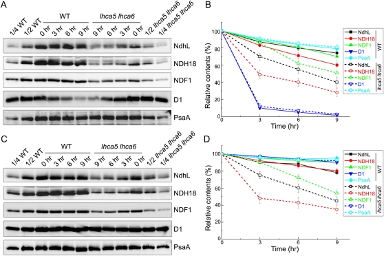
Figure 6.
Analysis of the stability of the NDH monomer and NDH-PSI supercomplex under high-light conditions. A, Leaves from 4-week-old wild-type (WT) and lhca5 lhca6 plants were vacuum infiltrated with lincomycin and cycloheximide for 30 min and then exposed to the high-intensity light (500 μmol photons m−2 s−1) for various times indicated at the top. After the illumination, thylakoids were isolated and subjected to SDS-PAGE and immunodetected with specific antibodies. B, Semiquantitative analysis of thylakoid proteins as in Figure 2B. The protein levels in the wild-type and lhca5 lhca6 leaves are shown relative to those in their leaves before high-light treatment (0 h; 100%). Values are means ± sd (n = 2). C and D, Conditions as in A and B, except that the leaves were vacuum infiltrated with water.

