Figure 1.
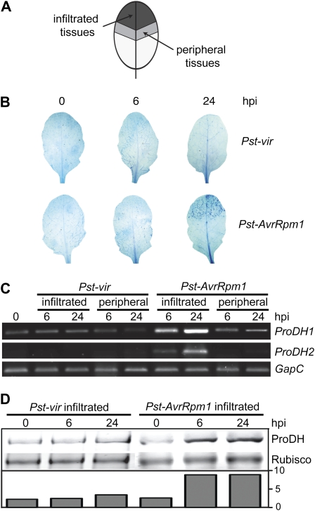
Early accumulation of ProDH at the center of the HR lesion. A, Sampling scheme. Suspensions of Pst-vir or Pst-AvrRpm1 isolates were locally inoculated (5 × 10⁶ cfu mL−1) at the ends of Arabidopsis leaves to then identify infiltrated and noninfiltrated peripheral tissues. B, Trypan blue staining of leaves excised at 6 or 24 hpi with pathogens. C, Quantification of ProDH1 and ProDH2 transcripts in pairs of samples (infiltrated, peripheral) isolated from pathogen-treated leaves as described in A. The assays involve semiquantitative, two-step, reverse transcription-PCR. The constitutively expressed GapC gene (At3g04120) was used as a control for cDNA content in the reactions. D, Western-blot analysis comparing the ProDH content in naive tissues and Pst-vir- or Pst-AvrRpm1-infiltrated tissues. The ProDH content is indicated at the bottom after normalization to Rubisco signal quantified by Ponceau S staining. The antibodies were generated against a peptide conserved between both ProDH isoforms, so that they may detect both proteins with similar sensitivity in these assays. One representative of three independent infection experiments is shown for each assay. Each experiment included six leaves (B) or four leaves (C and D) isolated from three different plants per time point. [See online article for color version of this figure.]

