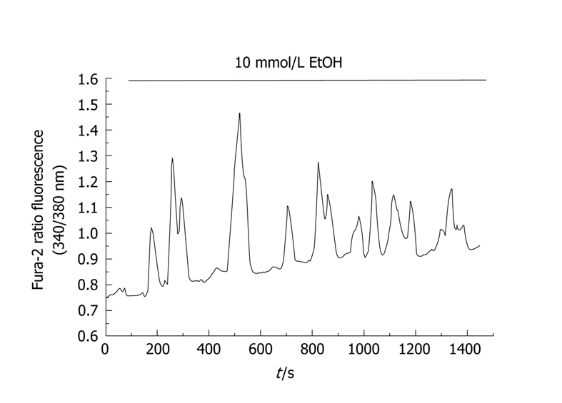
Figure 1.

Time-course of changes in [Ca2+]i in response to ethanol. Cells were loaded with the fluorescent probe fura-2. Changes in fluorescence emitted by the fluorophore reflect changes in [Ca2+]i. In this setup, pancreatic acinar cells were stimulated with 10 mmol/L ethanol, which induced an oscillatory pattern in [Ca2+]i. The horizontal bar indicates the time during which ethanol was applied to the cells. (nm, nanometers).
