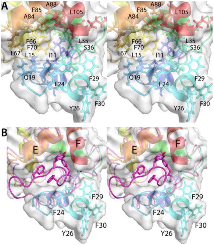
Fig. 6.

Structural changes accompanying removal of Ca2+ from ATH. (A) Stereoview of the hydrophobic cavity created by the movement of helix B. (B) This stereoview of the region of interest compares the path of the polypeptide backbone in the Ca2+-free and Ca2+-bound (magenta) states. Note that large displacements of the polypeptide chain are confined to the vicinity of the AB loop and the B helix.
