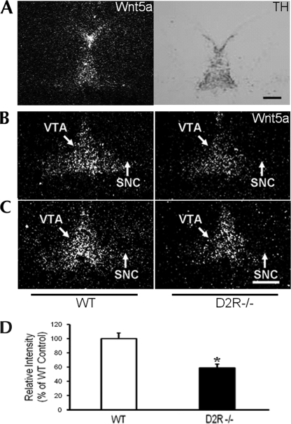
FIGURE 2.
In situ hybridization of Wnt5a gene expression in WT and D2R−/− mice. A, immunohistochemical image of TH staining and in situ hybridization image of Wnt5a in the mesencephalon of an E15.5 embryo brain are shown. B and C, In the middle (B) and end (C) of the SNC and VTA, expression levels of mesencephalic Wnt5a mRNA were assessed by in situ hybridization in WT and D2R−/− E14.5 mouse brains. Scale bars, 500 μm. D, levels of mRNA are expressed as percentage of WT controls. All values are expressed as means ± S.E. *, p < 0.05, n = 3 each.

