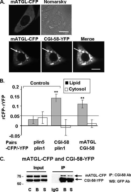
FIGURE 3.
CGI-58 interacts with mATGL (S47A) at the lipid droplet surface as determined by in situ AFRET and co-IP. Images were collected by confocal microscopy of live cells expressing pairs of fluorescent fusion proteins (A), and calculations for AFRET were performed for pairs of proteins indicated along the x axis (B). The gray line indicates the threshold of significance. Data are means ± S.E. from six to nine experiments. **, p < 0.01. C, CHO-K1 cells were co-transfected with mATGL(S47A)-CFP and CGI-58-YFP, and the cells were incubated with 400 μm oleic acid overnight. The following day, cells were incubated with either 5 μm triacsin C (B, basal conditions) or for 30 min with 10 μm forskolin, 1 mm IBMX, and 5 μm triacsin C (S, stimulated conditions). Cell lysates were incubated with rabbit anti-CGI-58 IgG (B and S) or rabbit preimmune control IgG (IgG). Immunoprecipitates were analyzed by Western blot (WB) for ATGL and CGI-58 using a commercial GFP antibody (Ab) that cross-reacts with YFP. One of three similar experiments is shown.

