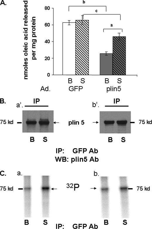
FIGURE 7.
Perilipin 5 effect on lipolysis and identification of perilipin 5 as a phosphorylated protein. A, AML12 cells were transduced with an adenoviral (Ad) construct for the expression of GFP or perilipin 5 (plin5) constructs for 48 h prior to lipolysis measurements. Cells were loaded overnight with [3H]oleic acid and 400 μm unlabeled oleic acid. Supplemental fatty acids were removed, and 5 μm triacsin C with (S) or without (B) 20 μm forskolin were added to the medium for 2 h. Release of fatty acids is shown for both basal (B) and stimulated (S) conditions. Data represent means ± S.E. (n = 16) (a, p < 0.01, for PKA stimulated value compared with basal value; b, p < 0.01; and c, p < 0.05 for cells expressing GFP compared with cells expressing perilipin 5. B and C, AML12 cells were transduced with adenoviral perilipin 5-YFP. Cells were incubated with 400 μm oleic acid overnight. 48 h after transduction, cells were loaded with [32P]orthophosphate for 2 h and then incubated for 30 min with (S) or without (B) 20 μm forskolin and 0.5 mm IBMX. Cell lysates were incubated with a commercial GFP antibody (Ab), and immunoprecipitates were analyzed by Western blot (WB) for perilipin 5. Two separate experiments were performed and are shown as indicated by panels a′ and b′ for Western blots (B), and panels a and b for autoradiograms (C).

