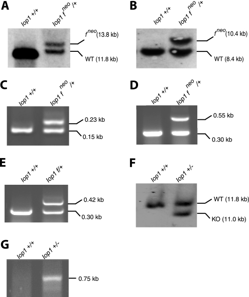
FIGURE 2.
Southern blotting and PCR confirmation of Iop1 gene targeting. A and B, Southern blots of ES cell DNA digested with HpaI (A) or BamHI/EcoRV (B) and hybridized with 5′ (A) or 3′ (B) Southern probes, respectively. The positions of the targeted allele (which contains the neomycin cassette) are indicated by fneo. C–E and G, PCR was performed on tail DNA to assess the presence of the 5′ loxP site (Iop LoxP5 and Iop LoxP3 primers) (C) and the neomycin cassette (Iop Neo and Iop Com primers; Iop Frt primer was also included to allow detection of the wild type allele at 0.30 kb) (D), the 3′ loxP site following deletion of the neomycin cassette (Iop Frt and Iop Com primers) (E), and the Iop1 knock-out allele (Iop Del and Iop Com primers) (G). The sizes of the PCR products are as indicated. Iop1 genotypes of the DNA samples are indicated above the gels. Iop1 f denotes the presence of the floxed allele in the absence of the neomycin cassette. F, Southern blot of tail DNA digested with HpaI and hybridized with the 5′ Southern probe.

