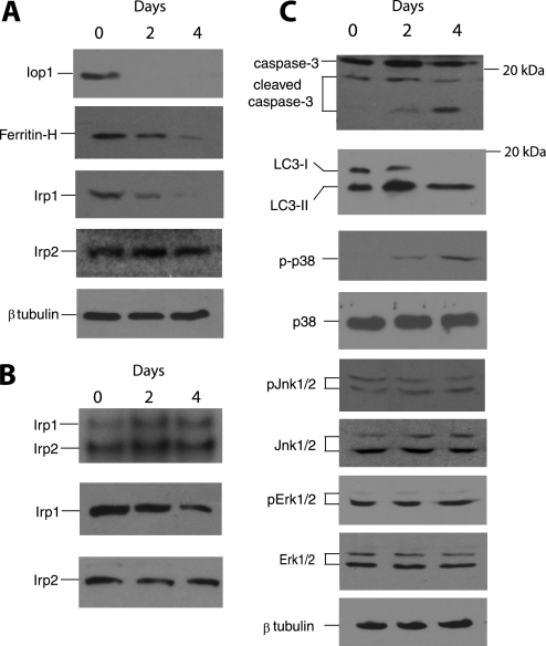
FIGURE 6.
Activation of selective intracellular pathways following Iop1 deletion in MEFs. A, Iop1f/f; Rosa26-creERT2 MEFs were treated with 4-hydroxytamoxifen for the indicated times. Cytosolic extracts were examined by Western blotting using the indicated antibodies. B, Iop1f/f; Rosa26-creERT2 MEFs were treated with 4-hydroxytamoxifen for the indicated times. Cytosolic extracts (20 μg) were incubated with a 32P-labeled RNA probe containing the ferritin heavy chain 5′ IRE and subjected to 5% PAGE (top panel). Irp1- and Irp2-induced gel mobility shifts are as indicated. Western blotting of extracts for Irp1 and Irp2 are shown in the bottom two panels. C, Iop1f/f; Rosa26-creERT2 MEFs were treated with 4-hydroxytamoxifen for the indicated times. Whole cell extracts (top two panels) or cytosolic extracts (all other panels) were examined by Western blotting using the indicated antibodies. The phosphorylated forms of p38, Jnk1/2, and Erk1/2 are denoted by p-p38, pJnk1/2, and pErk1/2, respectively. Positions of select molecular mass markers (in kDa) are indicated to the right of some panels.

