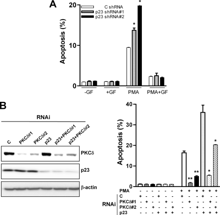
FIGURE 4.
The potentiation of PMA-induced apoptosis by p23 RNA depletion is mediated by PKCδ. A, LNCaP cells expressing p23 shRNA were treated with 30 nm PMA for 1 h either in the presence of absence of the PKC inhibitor GF 109203X (5 μm), which was added 1 h before and during PMA treatment. Apoptosis was assessed 24 h later. *, p < 0.05 versus control. B, LNCaP cells were transiently transfected with different RNAi duplexes, as indicated in the figure. Forty-eight h later, cells were treated for 1 h with 30 nm PMA (+PMA) or vehicle (−PMA), and apoptosis was assessed 24 h later (right panel). Expression of PKCδ and p23 was determined by Western blot (left panel). Results are expressed as mean ± S.D. of triplicate samples. *, p < 0.05; **, p < 0.01 versus control (C) RNAi. Similar results were observed in three independent experiments.

