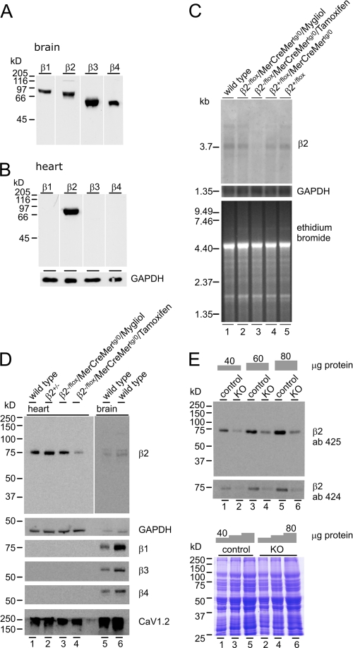
FIGURE 2.
Cavβ-expression. A and B, CaVβ1(β1), CaVβ2(β2), CaVβ3(β3), and CaVβ4(β4) in brain (A) and expression of Cavβ2 in heart (B) (100 μg/lane). C, CaVβ2 transcript expression in cardiac poly(A+)-RNA (10 μg/lane) from WT (lane 1), CaVβ2−/flox/MerCreMertg/0 (mock, lane 2), Tamoxifen-treated CaVβ2−/flox/MerCreMertg/0 (KO, lane 3), CaVβ2+/flox/MerCreMertg/0 (lane 4), and CaVβ2+/flox/MerCreMer0/0 (lane 5). Loading controls are as follows: GAPDH expression (middle) and ethidium bromide stain (bottom). D, CaVβ2 protein expression in heart (45 μg) from WT (lane 1), CaVβ2+/− (lane 2), CaVβ2−/flox/MerCreMertg/0 (mock, lane 3), and Tamoxifen-treated CaVβ2−/flox/MerCreMertg/0 (KO) (lane 4). Lane 5, 40 μg; lane 6, 80 μg. Protein expression in brain is shown as control (exposed for 15 min when compared with 1 min (lanes 1–4)). GAPDH was used as a loading control. E, estimation of the CaVβ2 protein expression levels in hearts from controls (lanes 1, 3, and 5) and KO (lanes 2, 4, and 6) using the CaVβ2 antibodies 425 (upper) and 424 (lower). Coomassie Brilliant Blue stain was used as a loading control.

