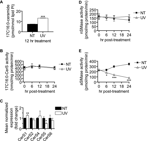
FIGURE 4.
UV-C irradiation increases CerS-derived Cer without up-regulating in vitro CerS activity or CerS expression or activating SMases. A, MCF-7 cells were treated (UV) or not treated (NT) with 10 mJ/cm2 UV-C followed by incubation for the indicated durations. Thirty minutes prior to harvest, 17C-Sph was added (1 μm). Following harvest, lipids were extracted, and 17C-Sph-containing ceramides were determined by HPLC/MS. Data represent mean and error bars represent S.E. of three independent experiments, and 17C-Cer levels were normalized to total lipid phosphate. Statistical significance was determined by Student's t test. B, cells were treated with UV-C as indicated, and whole cell lysates were prepared for the determination of in vitro CerS activity as described under “Experimental Procedures.” Data represent mean and error bars represent S.E. of four independent experiments assayed in duplicate. Statistical significance was determined by two-way ANOVA and Bonferroni post hoc tests. C, RT-quantitative PCR determination of CerS1–6 mRNA levels 12 h following UV-C irradiation. CerS3 was near or below detectable levels in all analyses. Data represent mean and S.E. of the -fold change above untreated cells. CerS mRNA levels were normalized to those of β-actin. Data represent three independent experiments, and statistical significance was determined by Student's t test. D and E, MCF-7 cells were treated with 10 mJ/cm2 UV-C followed by incubation for the indicated durations. Cells were then harvested, and nSMase (D) and aSMase (E) activities were determined as described under “Experimental Procedures.” Data are mean and error bars represent S.E. for three to five independent experiments. *, p < 0.05; **, p < 0.01; ***, p < 0.001 versus untreated control cells.

