FIGURE 7.
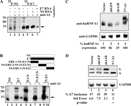
Binding and gene dosage analyses of hnRNP A1 in exon 7. A, unlabeled competitor RNA titrates hnRNP A1 association with exon 7 from nuclear extracts. Nuclear extract was incubated under splicing conditions with 0.28 pmol of uniformly labeled 32P-labeled NS RNA, 32P-labeled exon 7 RNA, and unlabeled competitor as indicated, treated with UV light, digested with RNase A, and then either directly resolved on a 12.5% SDS-PAGE gel (lanes 1 and 4) or immunoprecipitated with anti-hnRNP A1 (lanes 2, 3, 5, 6, and 7). B, hybrid NS and exon 7 RNA probes were used to narrow the hnRNP A1 binding site to the 3′ terminal end of exon 7. The schematic above the figure shows the NS RNA (white box), E7 RNA (black box), and the 20-nt ERE-1, ERE-2, and ERE-3 RNAs (black box) fused to NS RNA to make a 53-nt RNA molecule. C, depletion and overexpression of hnRNP A1 results in the increased production of CEACAM1-L and CEACAM1-S, respectively. Immunoblot analyses of HEK-293 cells using anti-hnRNP A1 is shown. D, RT-PCR analysis of mini-genes from C (sh-GFP is directed to GFP, sh-A1B is directed to hnRNP A1, and T7-A1 is over-expression) is shown. The mean ± S.E. for at least n = 3 is shown. Arrows denote cross-linking and/or immunoprecipitated hnRNP A1. **, p < 0.01 versus vector control.

