FIGURE 1.
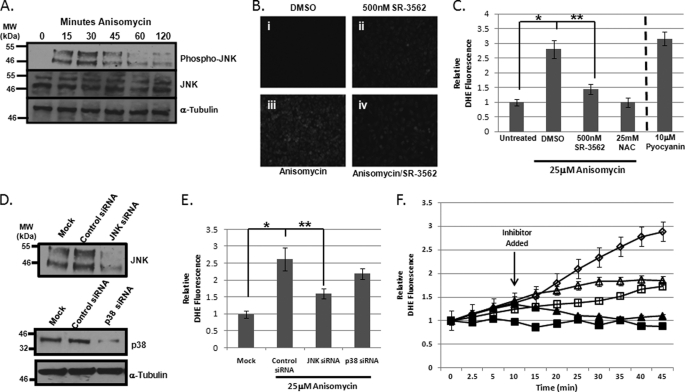
Anisomycin-induced stress initiates JNK-dependent ROS generation. A, JNK is activated by anisomycin in HeLa cells. JNK activation over the course of 2 h of 25 μm anisomycin stress was determined by Western blotting for bisphosphorylated JNK on Thr183 and Tyr185 (top). JNK abundance was monitored by Western blotting for total JNK (middle). α-Tubulin served as a loading control bottom. B, fluorescence microscopy of cellular ROS as detected by DHE. DMSO-treated cells (i) were compared with cells treated with 500 nm SR-3562 for 30 min (ii), cells stressed with 25 μm anisomycin for 45 min (iii), and cells pretreated for 30 min with 500 nm SR-3562 then stressed with 25 μm anisomycin (iv). C, fluorometric quantitation of cells stained with DHE to detect cellular ROS. Cells were stained with DHE following treatment with DMSO, 500 nm SR-3562 (30 min), 25 μm anisomycin (45 min), and 500 nm SR-3562 (30 min prior to anisomycin) and 25 μm anisomycin (45 min). 25 mm NAC, an antioxidant, was used as a negative control for ROS generation, and 10 μm pyocyanin, an inducer of ROS generation, was used as a positive control. D, Western blot analysis of JNK and p38 siRNA knockdowns of gene expression. Relative percent knockdown was estimated using densitometry. E, quantitation of cellular ROS in anisomycin-treated HeLa cells with JNK or p38 knockdown. After a 72-h siRNA transfection, cells were treated with 25 μm anisomycin for 45 min. ROS was detected using DHE staining and fluorescence was measured. F, kinetic profile of ROS generation in anisomycin-stressed HeLa cells. Cells were prestained with DHE prior to treatment with 25 mm NAC (black squares), 25 μm anisomycin (open diamonds), or 500 nm SR-3562 (open squares). Fluorescence was monitored for 45 min following the addition of the treatment. In two experiments, 25 mm NAC (black triangles) and 500 nm SR-3562 (open triangles) were added 10 min after 25 μm anisomycin.

