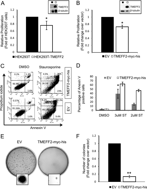
FIGURE 1.
TMEFF2 inhibits proliferation and anchorage-independent growth and sensitizes cells to apoptosis. A–F, stable expression of untagged (A) or c-Myc-His (B–F)-tagged form of TMEFF2 decreases proliferation of HEK293T cells (A and B), sensitizes the cell to an apoptotic stimulus (C and D), and inhibits anchorage-independent growth (E and F). Overexpression of TMEFF2 was confirmed by Western blot analysis (A and B, insets). The effect of TMEFF2 on growth (A and B) was determined using an MTT assay after 96 h of growth. The A562 at 96 h was normalized first to the value obtained at zero time (to correct for plating variability) and then to the value obtained for the parental cell line (HEK293T; A) or the cell line carrying the empty vector (EV) (B) as control. The effect of TMEFF2 on apoptosis of HEK293T cells (C and D) was determined in the presence of staurosporine or the vehicle, as a control, by analyzing the number of annexin V-positive cells and comparing it with the numbers obtained when expressing the empty vector. C and D, a representative image of the flow cytometry analysis (C) and percentage of apoptotic cells (D). E and F, a representative image showing anchorage-independent growth (E) and number of colonies formed by HEK293T cells stably expressing TMEFF2-Myc-His or the empty vector as a control (F) after 14 days of growth. Data shown are mean ± S.D. of at least three independent experiments with multiple replicates. Several clones were tested to rule out that the effects are due to the insertion site. *, p < 0.05, and **, p < 0.01.

