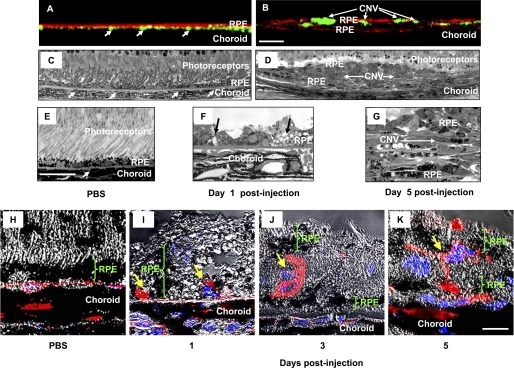
FIGURE 2.
Histological investigation of pathological changes after PEG-8 injection. A and B, X-view of the three-dimensional reconstruction of the RPE-choroid flat mounts of PBS- and PEG-8-treated animals, respectively. FITC-dextran-perfused vessels (arrow) are green, and the RPE cells stained with cytokeratin 18 are red. Subretinal tissue, including CNV, is located between two layers of the RPE cells (existing and newly growing). C–E and G, microphotographs of Epon-embedded semithin eye sections (1 μm) of PBS-injected mice (C and E) and PEG-8-injected mice (D and G) killed at day 5 post-injection. Subretinal tissue and CNV are located between two layers of the RPE cells, vessels containing erythrocytes marked with arrows. F, microphotograph of a semithin eye section of PEG-8-injected mice killed at day 1 post-injection showing the increased size of the RPE cells and vacuoles in the RPE cells (arrows). H–K, microphotographs of 5-μm paraffin sections of PBS-injected mice (H) and PEG-8-injected mice killed at day 1 (I), 3 (J), and 5 (K) post-injection. Isolectin IB4-labeled (red) endothelial cells penetrated the Bruch's membrane and growth between the RPE cells (I); endothelial cells proliferated in the subretinal space and formed new vessels partially covered by a new layer of RPE cells (J and K). The nuclei of cells were stained with DAPI (blue). Scale bars = 50 μm (A–D), 20 μm (E–G), and 10 μm (H–K).

