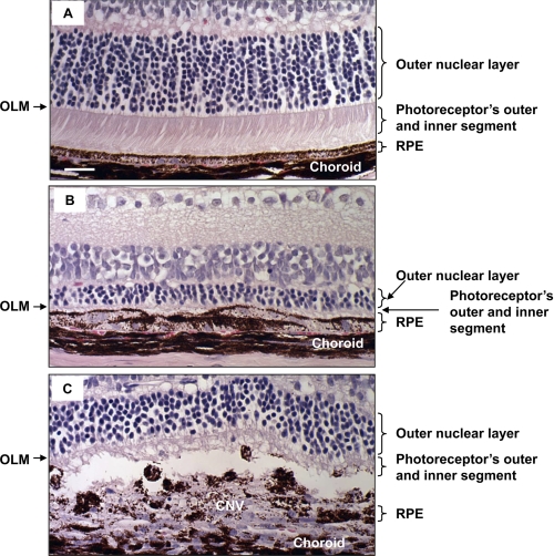
FIGURE 3.
Retinal changes after PEG-8 injection. Shown are microphotographs of paraffin sections of PBS-injected (A) and PEG-8-injected (B and C) animals killed at day 5 post-injection stained with hematoxylin and eosin. A decreased thickness of the outer nuclear layer and photoreceptor inner and outer segments was found in PEG-8-treated animals (B and C) compared with PBS-treated controls (A). OLM, outer limiting membrane. Scale bar = 20 μm.

