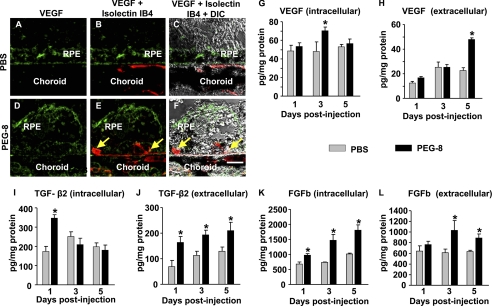
FIGURE 7.
Effect of PEG-8 on VEGF and other growth factors in the RPE-choroid. Microphotographs of paraffin sections show that PEG-8 increased VEGF expression on the apical side of the RPE and within the choroid (green; D–F) compared with the PBS-treated control (A–C). VEGF ELISA showed significantly increased levels of VEGF in the intracellular fraction of the RPE-choroid at day 3 after PEG treatment (G) and significantly increased levels of VEGF in the extracellular fraction of RPE-choroid at day 5 (H). TGF-β2 ELISA demonstrated significantly increased levels of TGF-β2 in the intracellular fraction at day 1 after PEG treatment (I) and significantly increased levels of TGF-β2 in the extracellular fraction of the RPE-choroid at days 1, 3, and 5 (J). bFGF (FGFb) ELISA showed significantly increased levels of bFGF in the intracellular fraction of the RPE-choroid at day 1, 3, and 5 after PEG treatment (K) and significantly increased levels of bFGF in the extracellular fraction of the RPE-choroid at days 3 and 5 (L). DIC, differential interference contrast. *, p < 0.05 compared with the PBS-treated group. Scale bar = 10 μm.

