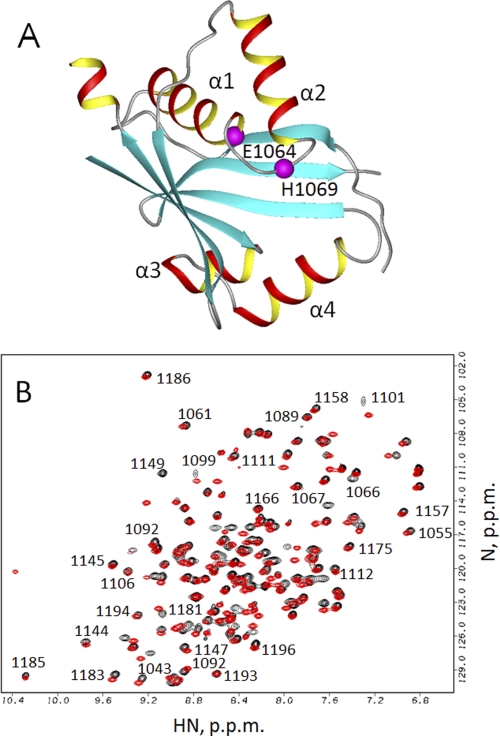
FIGURE 1.
A, structure of the wild-type ATP7B N-domain (Protein Data Bank ID code 2ARF). Residues His1069 and Glu1064 are shown in magenta. The unstructured loop (residues 1115–1138) is not shown. B, overlay of the 1H,15N HSQC spectra of the Glu1064-WNDΔ1115–1138 (red) and Ala1064-WNDΔ1115–1138 (black). Some of the sequential assignments for Ala1064-WNDΔ1115–1138 are shown in the uncrowded regions of the spectrum.

