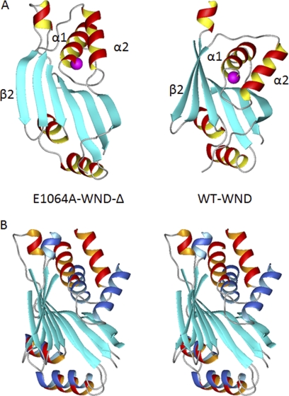
FIGURE 3.
Position of the α1–α2 hairpin differs between the wild-type WND and E1064A mutant. A, ribbon diagram of the E1064A-WNDΔ (left) and wild-type N-domain (right), with Glu/Ala1064 shown in magenta. B, stereo view of the wild-type N-domain (α-helices shown in blue) and the E1064A-WNDΔ (α-helices shown in red) aligned to minimize r.m.s.d. between the core β-sheets (cyan). The disordered loop in the wild-type N-domain is not shown (A and B).

