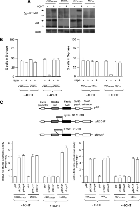
FIGURE 1.
Conditionally active AKT regulates rapamycin hypersensitivity and cyclin D1 and c-MYC IRES activities. A, immunoblot analysis of LN229 and MEF cells expressing the myr-AKT-MER fusion protein or EV control transfectants. The indicated cells were treated with the ligand 4OHT (1 μm, 24 h), and the extracts were prepared and separated by SDS-PAGE. The blots were incubated with anti-AKT, anti-Ser(P)473-AKT, and anti-β-actin antibodies. The black and gray arrowheads represent the myr-AKT-MER and endogenous AKT, respectively. B, LN229AKT-MER and LN229EV (left panel) and MEFAKT-MER and MEFEV (right panel) were treated in the presence or absence of 4OHT and rapamycin as shown and subjected to propidium iodide staining followed by flow cytometry. The means ± S.D. are shown for three independent experiments. C, AKT-dependent differential cyclin D1 and c-MYC IRES activities in myr-AKT-MER-expressing cell lines following 4OHT and rapamycin treatment as shown (LN229AKT-MER and LN229EV, left bottom panel; MEFAKT-MER and MEFEV, right bottom panel). The top panel depicts schematic diagrams of the dicistronic vectors. The relative fold change in firefly luciferase activity is shown as compared with activities obtained in the absence of rapamycin and normalized to values obtained for pRF in each cell line. The means ± S.D. are shown for three independent experiments.

