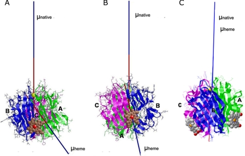
FIGURE 6.
Heme-induced alteration of the direction of the electric moment vectors of gC1q. The direction of the electric moment vectors (μ) two most probable gC1q-heme docking complexes are represented in a native form (μ native), as calculated from the Protein Data Bank code 1PK6 structure and in presence of heme (μ heme). The docking complexes where heme is bound to the A-chain (A-model, panel A) and to the C-chain (C-model, panel B), or both (panel C) are depicted. The three chains of gC1q are colored as follows: A, green; B, blue; and C, magenta. The heme is shown in sphere representation.

